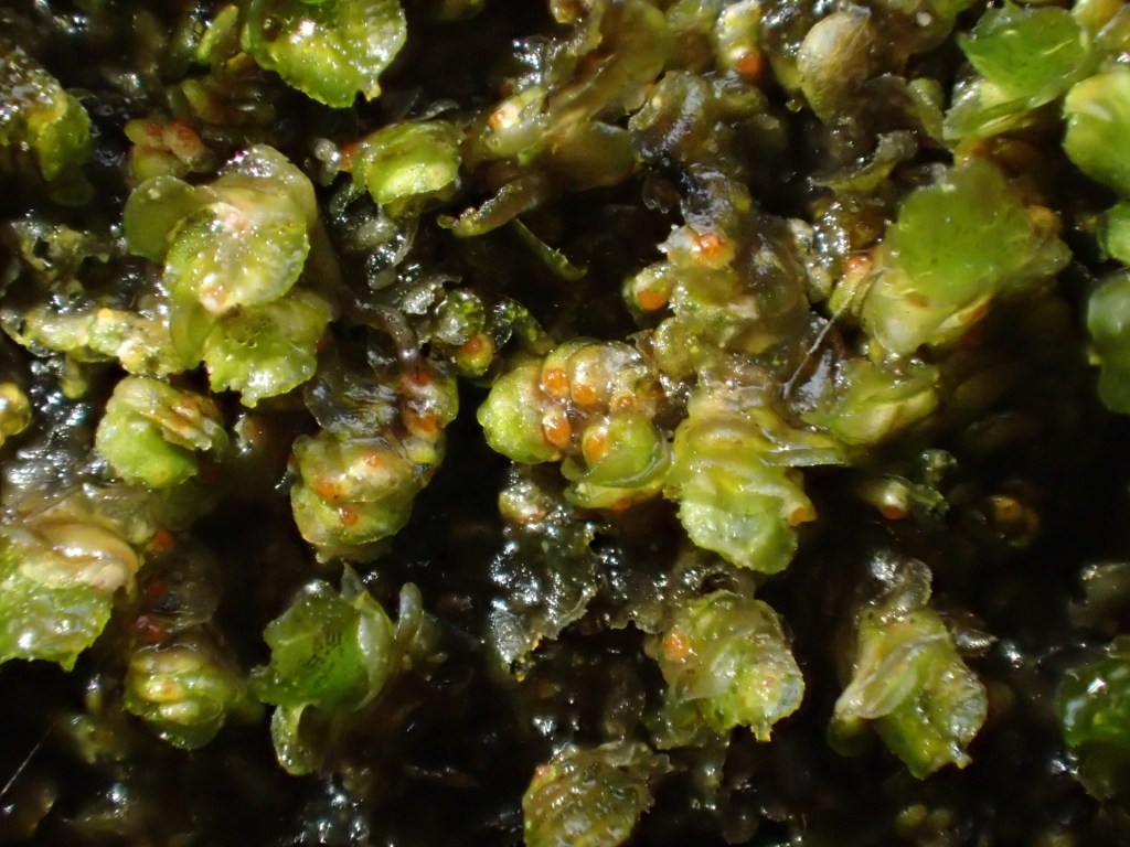
Hosts: Frullania dilatata
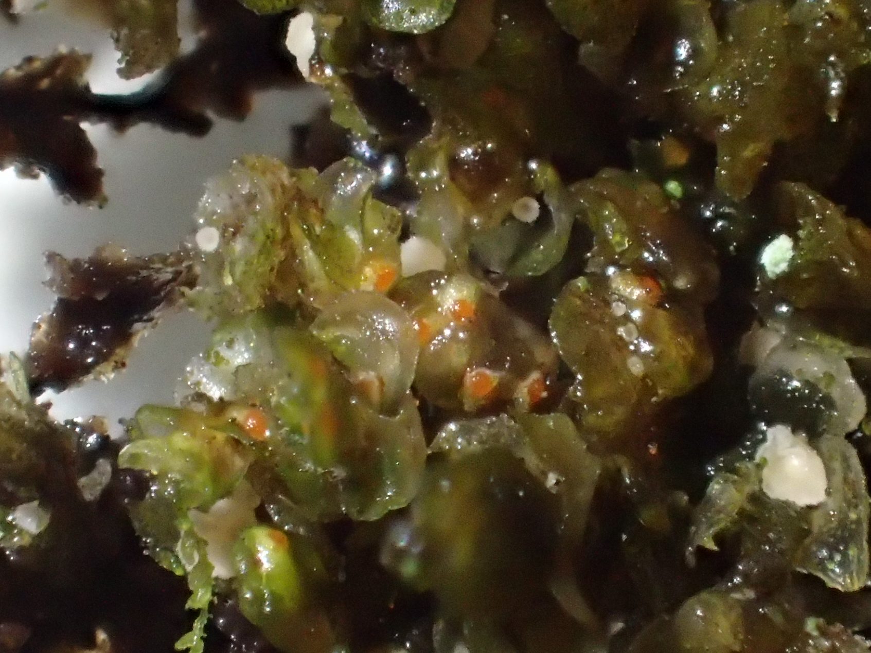
B. brongniartii is one of the commonest bryophilous ascomycetes found on Frullania dilatata, seemingly throughout the year. The globose, orange perithecia develop in the sheltered spaces beneath host lobes. When a perithecium is mature, the ostiole perforates through the host cell layer to disperse numerous minute ascospores. Infections are asymptomatic, but B. brongniartii may grow with necrotrophic species, like Pithyella chalaudii, that kill the host, turning the plants grey.

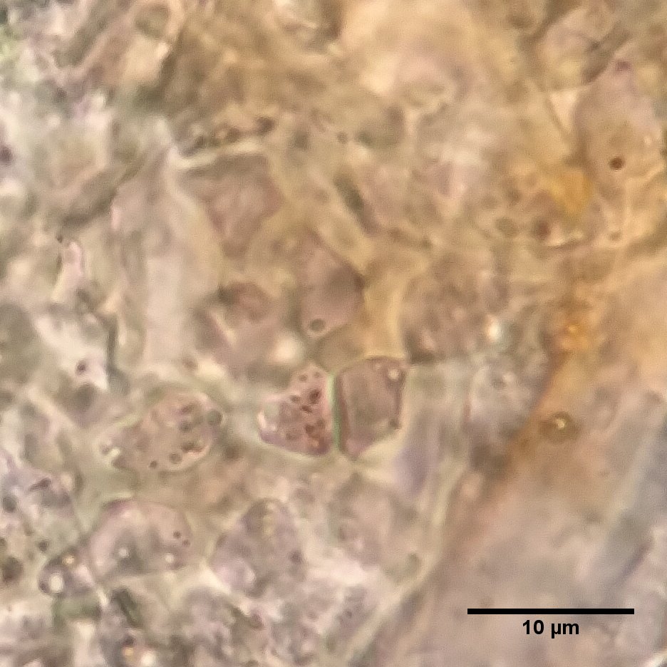
B. brongniartii is easily visible in the field under a hand lens as bright, smooth orange spots. The perithecia reach a maximum of about 1/4 of a millimetre in diameter (250 micrometres). Microscopically, the 8-spored asci and ascospores are very small and an oil immersion lens is recommended to appreciate them (though even 1000X total magnification is not powerful enough to satisfactorily visualise the microstructures of this fungus).
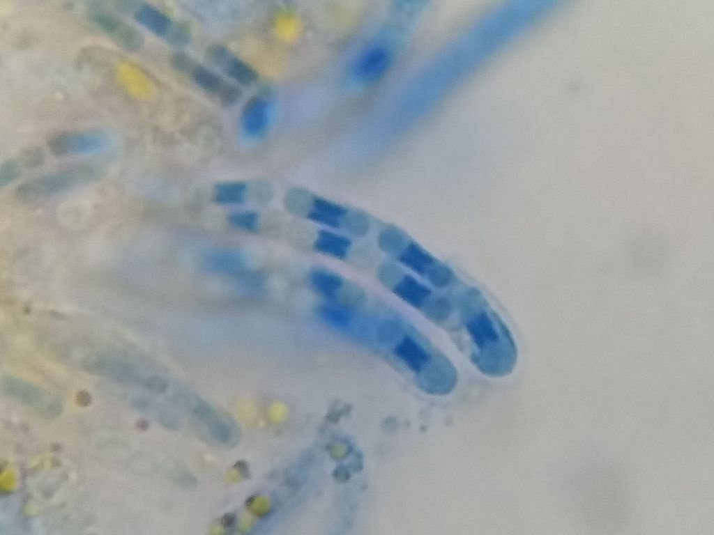
The ascospores are dumbbell-shaped and 1-septate, about 6-8μm long and 2-2.5μm wide. The septa, however, are frequently obscured by a distinctive, thickened structure in the medial domains of the spores (see images below). This band stains strongly in lactophenol cotton blue, so is therefore termed cyanophilous, and the variation in these bands is useful for separating species in the genus Bryocentria. Rupturing the fruitbodies of this fungus also releases of globules of orange pigment into the mounting medium. I have not noticed these in B. hypothallina and B. metzgeriae, only in this species and B. octosporelloides.
Notes on similar species:
B. brongniartii appears to be host specific. It has never been found on Frullania tamarisci or any other Frullania spp. in Europe (but has been in the USA), but it is worth keeping in mind that other Jubulaceae in the British Isles have not been screened much for fungal parasites. Our other Bryocentria species have different (principal) hosts, though in mainland Europe B. metzgeriae may occasionally colonise F. dilatata as well as the host it is more often found on, Radula complanata.
Bryocentria metzgeriae is separated from B. brongniartii based on two main features: (i) mode of parasitism – B. brongniartii is a biotrophic parasite that barely damages the host, while B. metzgeriae is necrotrophic and kills infected host patches (turns the thallus grey); (ii) spore structure – the spores of B. metzgeriae are smaller and spindle-shaped (fusiform) than the obviously dumb-bell shaped spores of B. brongniartii.
Octosporella erythrosigma is either rare, overlooked and / or highly seasonal in the British Isles, with only a single published record thus far. The perithecia of this fungus are also orange, but develop on the surface of the host lobes rather than emerging from beneath them, and they also possess obvious pale hairs (setae) on their surfaces. Microscopically, the aseptate, narrowly ellipsoid spores of O. erythrosigma are much larger than those of any Bryocentria.
Periantria frullaniae also forms smooth orange spots on F. dilatata, but these are strictly confined to the perianths of the host. Microscopically, the asci and spores of Periantria are massively elongated, the spore multiseptate. Vigorous infections of B. brongniartii may extent onto the perianths, so it is worth double-checking any fruitbodies found in them.
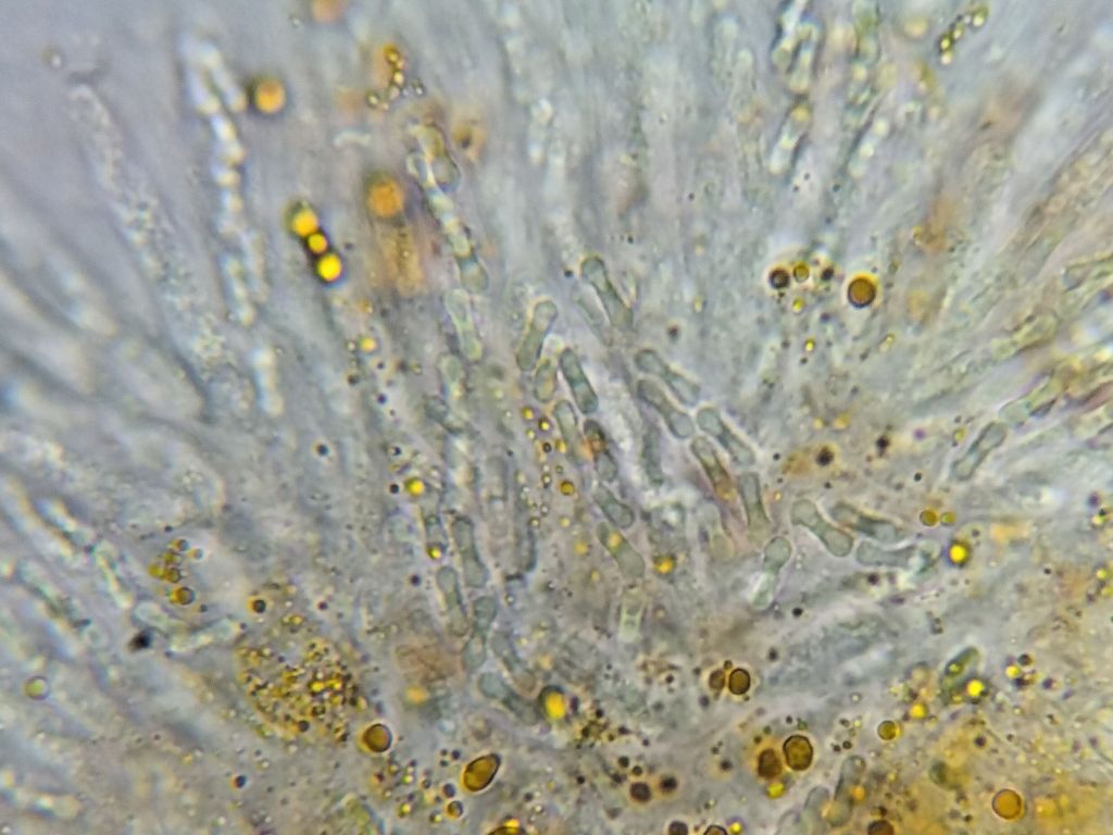
Trentepohlia algae frequently grow on bryophytes, looking somewhat like a rust fungus, especially on Frullania. These hairy orange balls may resemble Octosporella spp., and I have been foxed by spherical colonies of Trentepohlia a number of times. Close examination shows a lack of a fruitbody. Microscopically, the alga is made up of pigmented, thickened, filamentous cells. I should also mention that Trentepohlia is incredibly common, perhaps more so than any fungus on Frullania.
References
- Döbbeler, P. & Davison, P.G. 2017. Frullania as a hotspot for hypocrealean ascomycetes: ten new species from Southeastern North America. Nova Hedwigia 106(1–2): 209–256.
- Döbbeler, P. 1978. Moosbewohnende Ascomyceten I. Die pyrenocarpen, den Gametophyten besiedelnden Arten. Mitt. Bot. Staatssamml. München 14: 1–360.
- Döbbeler, P. 2002. Microniches occupied by bryophilous ascomycetes. Nova Hedwigia 75: 275–306.
- *** Döbbeler, P. 2004a. Bryocentria (Hypocreales), a new genus of bryophilous Ascomycetes. Mycological Progress 3: 247–256.
- Döbbeler, P. 2004b. Octosporella erythrostigma (Pezizales) and Pithyella frullaniae (Helotiales), two remarkable ascomycetes on Frullania dilatata. Feddes Repertorium 115: 5–14.
- *** Döbbeler, P. 2006. Ascomycetes on Frullania dilatata (Hepaticae) from Tuscany. Mycol. Progress 5: 32–40.
- Döbbeler, P. 2010. New species and records of Bryocentria – a hypocrealean genus of bryophilous ascomycetes. Karstenia 50: 11–23.
- Döbbeler, P., Linde J. & E. Rubio. 2018. Bryocentria octosporelloides (Hypocreales) – a new species on Cololejeunea minutissima from Asturias (Spain). Ascomycete.org 10(2): 77–80.
- Greiff, G.R.L. 2021. Studying bryophilous fungi on Frullania dilatata. Field Bryology 126: 35–40.
- Nordén B., Gardiennet A., Priou J.-P. & Döbbeler P. 2015. Bryocentria hypothallina (Hypocreales) – a new species on Metzgeria furcata. Ascomycete.org. 7(4): 121 – 124.
- Priou, J.-P. 2019 Pithyella chalaudii Priou sp. nov. (Helotiales) pour remplacer P. frullaniae (Chalaud) Döbbeler inval. Ascomycete.org: 11(3).
- Racovitza, A. 1959.Étude systématique et biologique des champignons bryophiles. Mémoires du Muséum National d’Histoire Naturelle, Série B, Botanique 10(1): 1–288.
- Stenroos, S., Laukka, T., Huhtinen, S., Döbbeler, P., Myllys, L., Syrjänen, K. & Hyvönen, J. 2010. Multiple origins of symbioses between ascomycetes and bryophytes suggested by a five-gene phylogeny. Cladistics 26: 281–300.
