Hosts: Rhizomnium punctatum


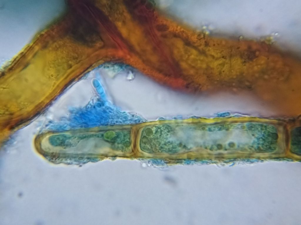


Remarks: This was an interesting collection found while attempting to visualise the haustoria of suspected Octospora mnii on the same host. It seems to be rather specialised, colonising only the chloronemal cells (cell walls at right angles to the growth direction rather than oblique – see image 1), particularly tip-growing cells. Infected tip cells appear to die, suggested by shrinkage and deformation, but their walls are naturally thin and those apparently free of fungi also shrink and have cytoplasmic aberrations. Water mounts of fresh collections are required to learn more about this fungus, determine how widespread it is, and whether it is a common associate of Rhizomnium punctatum.
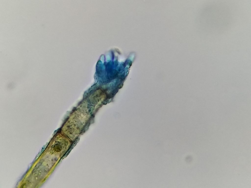
Initially, I suspected that the conidia to be protonemal gemmae, however the surrounding mycelium and morphology of the conidia point towards a fungus. I am under the impression that protonemal gemmae are usually think-walled and often contain brown pigment, and that they do not form in bunches. But I have little experience in this regard and will continue to examine the protonema of Rhizomnium for answers.
